Background: Cerebellopontine angle lesions represent a
significant proportion of intracranial tumors, with schwannomas comprising the
majority. Magnetic resonance imaging has revolutionized the diagnostic approach
to these lesions with its superior soft tissue contrast resolution and
multiplanar capabilities.
Objective: To evaluate the role of magnetic resonance
imaging in localizing and characterizing cerebellopontine angle schwannomas and
to compare findings with surgical and histopathological outcomes.
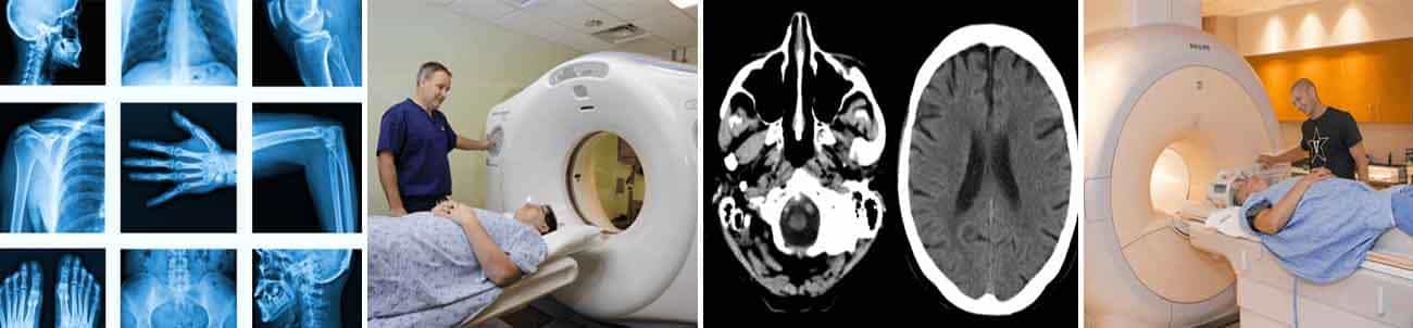
Methods: A prospective observational study was
conducted over five months involving 20 patients presenting with clinical
features suggestive of cerebellopontine angle lesions. All patients underwent
magnetic resonance imaging using a 1.5 Tesla Phillips scanner with standard
protocol including T1-weighted, T2-weighted, diffusion-weighted imaging, and
fluid-attenuated inversion recovery sequences in axial, sagittal, and coronal
planes. Gadopentate dimeglumine was administered as contrast agent where
indicated. Imaging findings were correlated with surgical and histopathological
results.
Results: The study population consisted predominantly
of patients aged 21-40 years, with female preponderance. Vestibular schwannomas
constituted 95% of cases, with vestibulocochlear nerve involvement in 85% of
patients. On T1-weighted images, 95% of schwannomas were hypointense relative
to brain parenchyma, while all cases demonstrated hyperintense signal on
fluid-attenuated inversion recovery sequences without restricted diffusion.
Post-contrast imaging revealed marked to moderate enhancement in all
schwannomas. Magnetic resonance imaging demonstrated 100% sensitivity, 92.86%
specificity, 94.12% positive predictive value, and 96.67% overall accuracy in
diagnosing vestibular schwannomas when correlated with histopathology.
Please enter the email address corresponding to this article submission to download your certificate.