ARCHIVES
VOL. 7, ISSUE 2 (2025)
Pediatric Upper Extremity Trauma Imaging: Building Blocks for the Developing Radiologist
Authors
Dr. Shrinivas Radder, Dr. Nivedita Radder
Abstract
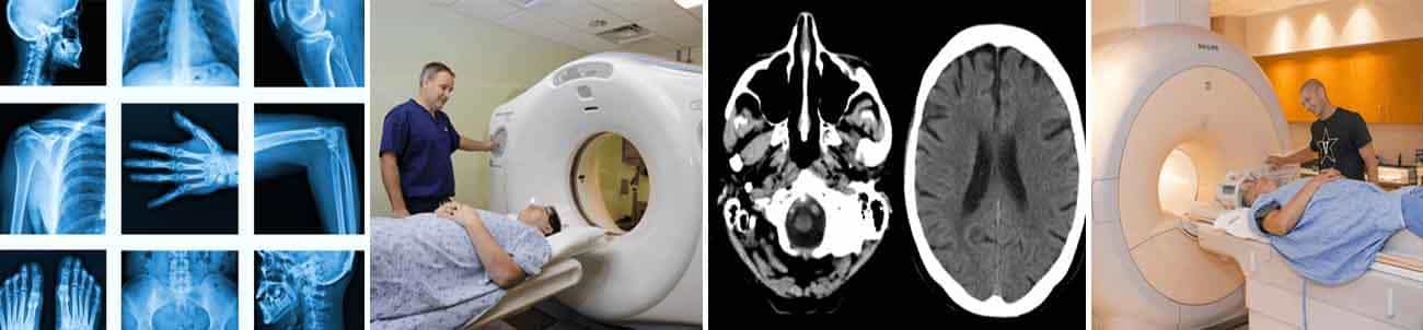
Pediatric upper extremity injuries are among
the most common reasons for emergency department visits in children, often
resulting from falls, sports activities, or non-accidental trauma. The unique
anatomical and physiological characteristics of the growing skeleton, including
the presence of open growth plates, secondary ossification centers, and
developing cartilage, present both diagnostic challenges and opportunities for
radiologists. Accurate imaging evaluation is crucial not only for identifying
fractures but also for recognizing subtle patterns that may indicate more
serious pathology, such as physeal injuries or child abuse. Radiographs remain
the first-line imaging modality, but advanced techniques such as ultrasound and
MRI play an increasingly important role in evaluating soft tissue injuries,
growth plate disruptions, and occult fractures. The use of low-dose CT and
tailored protocols further aids diagnosis while minimizing radiation exposure.
Understanding the normal developmental anatomy, especially in complex regions
like the elbow where multiple ossification centers appear in a predictable
sequence is essential to avoid misinterpretation. Furthermore, recognizing
age-specific injury patterns such as buckle fractures, supracondylar fractures,
and Monteggia variants is critical for accurate reporting and appropriate
management. Classification systems like Salter-Harris remain indispensable
tools for describing fracture patterns involving the physis. The radiologist
must also remain alert to red flags for non-accidental trauma, particularly in
infants and toddlers, where imaging findings may be the first or only indicator
of abuse. This article provides a comprehensive review of imaging strategies,
developmental considerations, common injury patterns, classification systems,
complications, and the evolving role of radiologists in the multidisciplinary
care of pediatric patients with upper extremity injuries. Emphasis is placed on
the integration of anatomical knowledge, imaging acumen, and clinical context
to ensure precise diagnosis and optimal outcomes for pediatric patients.
Download
Pages:31-38
How to cite this article:
Dr. Shrinivas Radder, Dr. Nivedita Radder "Pediatric Upper Extremity Trauma Imaging: Building Blocks for the Developing Radiologist". International Journal of Radiology Research, Vol 7, Issue 2, 2025, Pages 31-38
Download Author Certificate
Please enter the email address corresponding to this article submission to download your certificate.