ARCHIVES
VOL. 1, ISSUE 4 (2019)
Computed tomography (CT) perfusion evaluation of brain lesions
Authors
Dayanand Kawade, Samruddhi Sonwane, Ashish Lule
Abstract
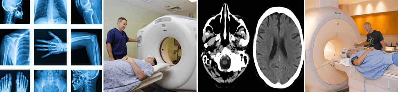
Introduction: Computed tomography perfusion (CTP) is a better choice for assessing hemodynamic parameters compared to Magnetic Resonance Imaging (MRI). With this perspective this study is undertaken to study perfusion characters and difference Material Method: 100 patients (64 Males and 36 Females) with age between 17 to 68 years include. Computed tomography perfusion (CTP) was performed with Dual‑Slice Dynamic Multi‑Detector Scanner. Result: perfusion parameters of lymphomas, low‑grade glioma, high‑grade gliomas, Grade 3 glioma and glioblastoma multiforme (GBM) were not found statistically significant. There were statistically significant differences in rCBF for lymphomas, low‑grade glioma, Grade 3 glioma, high‑grade gliomas and glioblastoma multiforme (GBM) in Lesional and PeriLesional area. Discussion: low perfusion in low‑grade gliomas, glioblastoma multiforme (GBM) is found. It may be because of tumor heterogeneity, necrosis and vascular invasion Conclusion: CT perfusion can be a good aid in differentiating various lesions.
Download
Pages:17-20
How to cite this article:
Dayanand Kawade, Samruddhi Sonwane, Ashish Lule "Computed tomography (CT) perfusion evaluation of brain lesions". International Journal of Radiology Research, Vol 1, Issue 4, 2019, Pages 17-20
Download Author Certificate
Please enter the email address corresponding to this article submission to download your certificate.